
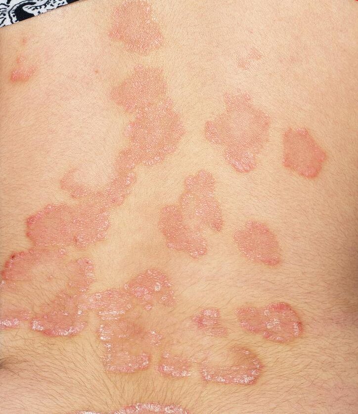
Glen is a 48-year-old IT professional who presents with a two-week history of painless scaly erythematous plaques on the lower back (pictured). He has psoriasis, which has been stable on monthly subcutaneous secukinumab 300mg for several years.
Glen was treated with antivirals for shingles in the same distribution one month ago. The blisters dried and crusted off over 10 days, and two days later, red lesions with white scale developed, overlying the healing zoster lesions. He has no associated neuralgia.
The lesions are confined to the area pictured. Dermoscopically, diffusely distributed white scales on a bright red background pattern are seen, as well as regularly arranged dotted globules and hairpin-like vessels.
What is the most likely diagnosis?
Correct!
Post-herpetic psoriasis manifests with unilateral scaly erythematous plaques at the location of previously healed herpes zoster. It can be distinguished from typical psoriasis by the distribution of lesions along the zoster-affected dermatomes.
The lesions in post-herpetic psoriasis are characterised by erythematous plaques with white scale and punctate bleeding when the scale is scraped away. The typical dermotoscopic findings are diffuse white scales over a bright-red background, with a regular distribution of dotted, annular and hairpin-like vessels.
This is an example of Wolf’s isotopic response, which describes the emergence of a new dermatosis at the site of a previous unrelated dermatosis (in this case, psoriasis at zoster site).
The other listed diagnoses may also present with Wolf’s isotopic response following herpes zoster. Zosteriform pityriasis rosea presents as a herald patch lesion with unilateral development of lesions at the prior zoster site. In such cases, dermoscopy reveals focal punctate or linear vessels on a yellow background with marginal scale distribution.
Lichen planus at the site of a healed herpes zoster manifests with flat-topped erythematous–violaceous polygonal papules with Wickham’s striae. These lesions present as a band along the course of a peripheral cutaneous nerve and its branches. Dermoscopically, there are white reticulate streaks and vessels are peripherally located.
A cutaneous lupus erythematosus Wolf’s isotopic response presents as pink scaly papules in the zoster-affected areas. Removal of the adherent scale results in visible keratinous follicular plugs, which are evident dermoscopically, along with perifollicular white haloes.
Post-herpetic psoriasis is managed, and typically resolves, with treatment for typical psoriasis. For mild manifestations, topical therapy, such as calcipotriol, may be sufficient. Those who are already on immunodulatory treatment can be continued on their regular treatment with surveillance.