
A 68-year-old Caucasian woman presents with an eight-year history of an erythematous, pruritic and scaly rash over her central chest, upper back and forearms.
This is on a background of breast cancer, which was managed with lumpectomy and radiotherapy, resulting in significant post-radiation dermatitis on her right breast.
She also has type 2 diabetes, hypertension, asthma and hypercholesterolaemia, which are managed with metformin, glimepiride, exenatide, perindopril, fluticasone propionate, salmeterol, salbutamol and rosuvastatin.
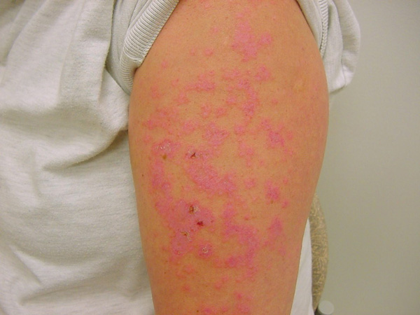
On examination, there are well demarcated erythematous macules and erosions on her upper chest (pictured), upper back and forearms. She has tried clotrimazole for two months with no improvement.
What is the most likely diagnosis?
Correct!
Discoid lupus erythematosus (DLE) is the most prevalent form of cutaneous lupus erythematosus.
It is a chronic inflammatory disorder initially characterised by erythematous macules and papules that typically evolve into disc-shaped scaly plaques. The rash is often asymptomatic, but can be pruritic and sore.
DLE is mostly localised (above the neck – 80%), but can be generalised (below the neck – 20%). It may progress into areas of hyperpigmentation with central atrophic scarring alongside hypopigmentation and telangiectasia. Scarring alopecia is common in affected hair-bearing areas.
DLE may occur due to endogenous factors, such as genetic predisposition, and may be aggravated by exogenous factors, including smoking and UV light exposure. It is more likely to occur on sun-exposed areas. It is most commonly seen in women aged 20-40, although both genders may be affected at any age.
The condition is becoming increasingly common and severe in people of colour, especially Indigenous Australians and those of South-East Asian descent. In people of colour, hyper- and hypopigmentary change is more pronounced.
Diagnosis is based on clinical examination, lesional skin biopsy and blood tests, including full blood count, kidney function, antinuclear antibodies (ANA), and extractable nuclear antigen (ENA). ANA and ENA are negative in 85% of patients with DLE, unlike subacute and systemic lupus erythematous, in which these markers are more likely to be positive.
Treatment includes photoprotection using sun-protective clothing, hats and sunscreen. Limited disease may be treated with topical corticosteroids or calcineurin inhibitors, with intralesional corticosteroid injection an option for escalation.
More extensive or treatment-resistant disease warrants dermatology input and may require systemic therapy with hydroxychloroquine, immune response modifiers (such as sulfasalazine or biologics) or immunosuppressants (such as systemic steroids, azathioprine or methotrexate).
For patients under consideration for hydroxychloroquine therapy, it is recommended to refer for screening for retinopathy (due to increased risk of this with higher hydroxychloroquine doses) and G6PD deficiency (in which case this agent is contraindicated).
Atopic dermatitis typically affects flexures. Dermatophytosis is less likely given the lack of improvement with antifungal therapy, and lack of characteristic ring-shaped lesions. Cutaneous metastatic breast cancer classically presents as firm, rubbery nodules over the breast and ipsilateral chest wall.