
Cherie is a 42-year-old physiotherapist who presents with a week of sudden onset of large red to brown skin lesions, associated with some blistering, on the lower limbs. These are itchy and burning at times.
The lesions initially appeared on her right foot, then progressed to her right thigh. Four days before the rash developed, Cherie had completed a course of norfloxacin for travellers’ diarrhoea, following a recent holiday in South-East Asia.
Apart from the rash, she feels well and the gastroenteritis has resolved. She has no medical history of note, no known allergies, and takes no medications.
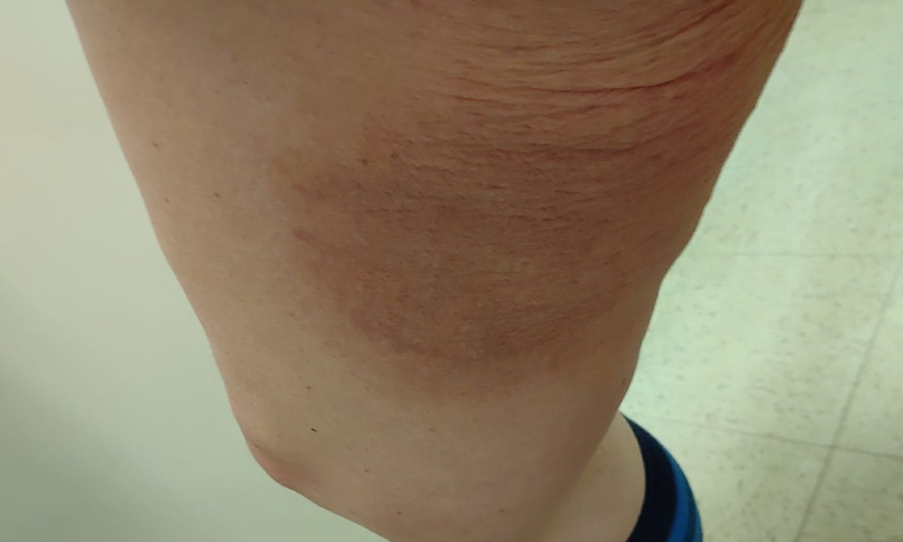
On examination, vital signs are normal. There are multiple hyperpigmented, non-tender, non-indurated macular lesions on the right leg and thigh (pictured).
What is the most likely diagnosis?
Correct!
The history is most consistent with a bullous fixed drug eruption (FDE) secondary to quinolone use. Cutaneous reactions affect 2-3% of patients taking medication, though exanthematous eruptions are more common than FDEs, which have an estimated prevalence of 0.003%.
FDE is a delayed type IV hypersensitivity reaction. Oral antimicrobials and NSAIDs are the most commonly implicated agents. Cases have also been reported associated with paracetamol, carbamazepine, benzodiazepines, calcium channel blockers, ACE-I, omeprazole and following vaccinations.
FDEs may be localised or generalised, though tend to be solitary or occur in small groups. They appear as well-demarcated, dusky red to brown lesions, but can present with varying morphology, including bullous, linear and non-pigmented lesions. These may be otherwise asymptomatic or be associated with pain or itch.
Lesions heal with scarring or hyperpigmentation and may recur at the same site on re-exposure. Unlike other drug reactions, the patient is systemically well.
Diagnosis is usually clinical, with a skin biopsy indicated in cases of diagnostic uncertainty. Management involves withdrawal of the offending agent followed by avoidance in future. Topical steroids and antihistamines may be of benefit for those who are symptomatic.
Immunobullous diseases, such as bullous pemphigoid and Stevens-Johnson syndrome, typically present with extensive involvement and systemic features. The non-dermatomal distribution and pattern of the rash are not consistent with typical herpes zoster.