
Aaron is a 50-year-old builder who presents after dropping a marble benchtop on the tip of his right middle finger. He reports that the finger swelled, “hurt like crazy and looked wrong” immediately after the injury.
Aaron is generally well, with well-controlled hypertension on candesartan. He has no other past history of note.
On examination, the affected distal interphalangeal joint is in a flexed position (pictured), with full passive extension but limited active extension. There is a subungual haematoma but no obvious nail bed injury.
What is the most likely diagnosis?
Correct!
Mallet finger occurs following disruption of the extensor digitorum tendon attachment to the dorsal distal phalanx.
Bony mallet finger describes an avulsion fracture of the base of the distal phalanx, with the extensor tendon remaining attached to the fracture fragment, compared with a soft mallet finger, which refers to an extensor tendon injury in isolation.
The most common cause is a sudden forced flexion injury to an extended distal interphalangeal joint (DIPJ) — for example, a ball to the tip of an outstretched finger — although lacerations and crush injuries can also disrupt the extensor tendon insertion.
The characteristic features of this injury are a resting fixed flexed position of the affected DIPJ and inability to actively extend the affected DIPJ (extensor lag).
There may also be compensatory proximal interphalangeal joint (PIPJ) hyperextension (swan-neck deformity).
Bony mallets generally heal more reliably than their soft tissue counterparts, and in most cases, both injuries can be successfully managed with an appropriately fitted thermoplastic splint.
This may be overseen by a suitably qualified hand therapist, who will create the splint and monitor for secondary consequences, such as hyperextension at the PIPJ. This hyperextension is a compensatory motion for the hyperflexion of the DIPJ.
The managing hand therapist will ensure flexion at the PIPJ rather than continuing the hyperextension of the PIPJ to offset the distal injury.
A bony mallet that involves the joint and is associated with a subluxed joint is a rare variant that does require surgery.
Tuft fractures involve the tip of the distal phalanx — typically following a crush injury. There is often an associated nail bed injury, indicating an open fracture.
Jersey finger is an avulsion injury of the flexor digitorum profundus from its insertion at the base of the distal phalanx. It usually follows a hyperextension injury to the phalanx and earned its name because a common mechanism of injury is a footballer grabbing the jersey of an opposing player.
Clinically, the affected finger rests in slight extension at the DIPJ.
A Seymour fracture is an important subtype of bony mallet finger occurring in children. It describes a distal phalanx physeal fracture with an associated nail bed injury — commonly with ungual subluxation.
These often occur in crush injuries and present with apparent mallet finger deformity with soft tissue trauma at the proximal nail fold.
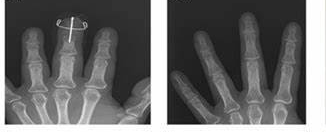
In this case, an X-ray confirms bony mallet finger. Aaron is referred to a hand therapist and fitted with a thermoplastic mallet splint for 6-8 weeks, with an uncomplicated recovery.