
Gerard is a 62-year-old retired architect who presents with 24 hours of a painful red right eye.
He describes deep aching pain in the eye and right temporal region that is worse with eye movement, in bright light and when reading. His vision is unaffected.
Gerard is generally well, with a history of hypertension treated with ramipril, and ulcerative colitis, which has been in remission for five years.
He has no recent URTI history, no sick contacts and denies foreign body exposure.
On examination, VA is 6/9 uncorrected bilaterally. The right pupil appears constricted and there is direct and consensual photophobia.
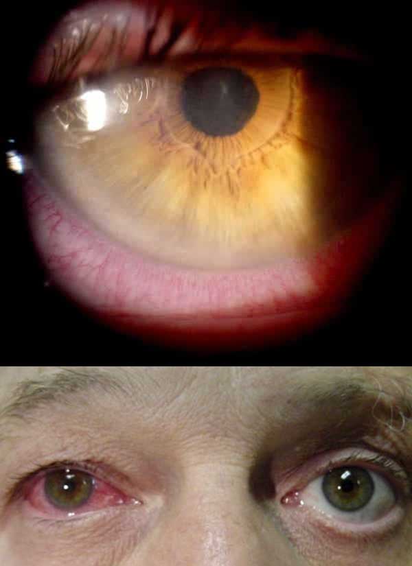
The right conjunctiva is erythematous; this is most prominent adjacent to the corneal limbus (pictured).
Fluorescein examination is unremarkable. The left eye examines normally.
What is the most likely diagnosis?
Correct!
The images demonstrate a constricted slightly irregular right pupil with ciliary flush (that is, circumcorneal involvement of the diffusely injected conjunctiva), consistent with uveitis.
While this may occur idiopathically, uveitis most frequently occurs in association with systemic conditions, particularly inflammatory diseases and infection.
Associated inflammatory conditions include the spondyloarthropathies, sarcoidosis, psoriatic arthritis, IBD and MS.
HLA B27 antigen is present in up to 70% of those with anterior uveitis.
Infectious causes include herpes virus, cytomegalovirus, toxoplasmosis, syphilis and tuberculosis. Rare cases have been reported with severe COVID-19 infection and post-COVID vaccination.
Drug-related uveitis is less common; rifabutin is a key potential trigger, with cases also occasionally seen with bisphosphonate therapy (typically IV) and BRAF-inhibitor therapy for metastatic melanoma.
The clinical presentation depends on the portion of the uveal tract involved.
Anterior uveitis is about four times more common than posterior uveitis, and typically presents with pain, often described as a deep ache radiating to the periorbital and temporal region and worse with movement and accommodation.
Redness is typically most marked at the limbus (ciliary flush), with a constricted pupil which may be irregular.
Posterior uveitis is more likely to be painless, and may cause nonspecific visual changes such as floaters and/or reduced visual acuity.
Redness is not a prominent feature unless there is an accompanying anterior uveitis.
Prompt ophthalmological assessment is warranted in suspected cases, for definitive diagnosis and assessment for potential complications, such as posterior synechiae (adhesion of the iris to the lens), intraocular hypertension, glaucoma and cataract.
Treatment is typically with topical corticosteroids, with topical dilating drops short term for pain relief.
Systemic steroids or steroid-sparing immunosuppressants may be required for recalcitrant cases.