Dermatoscopy, also known as dermoscopy, is a non-invasive diagnostic tool that allows for the visualization of skin lesions with greater clarity and magnification. It’s particularly useful in distinguishing benign from malignant skin lesions. Here’s an overview of dermatoscopic findings in common skin lesions:
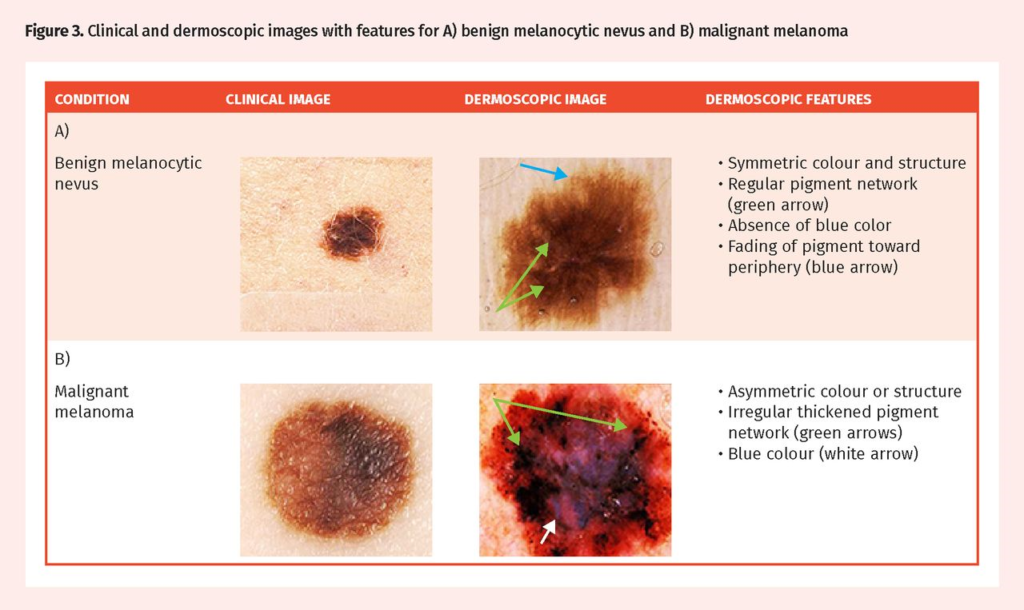
- Melanocytic Nevus (Mole):
- Regular, symmetric pattern.
- Uniform color, typically tan, brown, or black.
- Homogeneous pigmentation or a reticular pattern (network-like structures).
- Presence of a pigment network that is evenly distributed and consistent in color and thickness.
- Melanoma:
- Asymmetry in structure and color.
- Irregular and uneven pigment network.
- Variability in colors (brown, black, gray, blue, red, white).
- Irregular dots or globules, streaks.
- Regression structures such as scar-like areas, or blue gray veil areas.
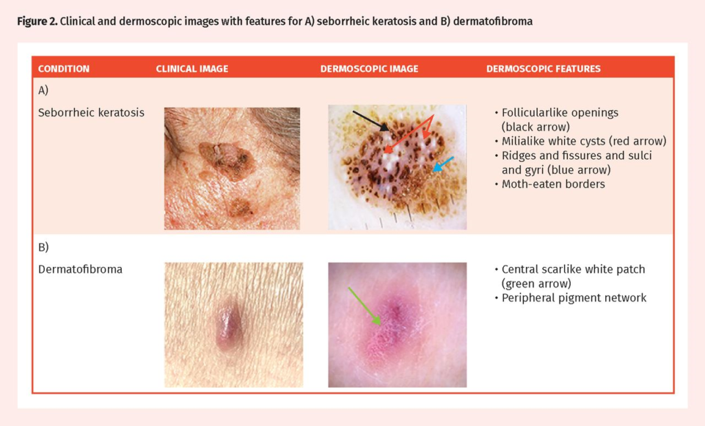
- Seborrheic Keratosis:
- Warty or stuck-on appearance.
- Milky-white, yellow, or light brown coloration.
- Comedo-like openings (black dots) and keratin cysts.
- A network of fine superficial vessels may be observed.
- Actinic Keratosis:
- Erythematous background with a scaly surface.
- Fine, superficial white scales.
- “Strawberry pattern” characterized by a red pseudonetwork with prominent follicular openings.
- May show small, dotted vessels.
- Squamous Cell Carcinoma (SCC):
- Hyperkeratosis (thickened, white scale).
- Irregular, coiled, or glomerular vessels.
- Erosions or ulceration.
- A yellow-white color in the background due to keratin.
- Basal Cell Carcinoma (BCC):
- Arborizing vessels (branching blood vessels).
- Blue-gray ovoid nests or globules.
- Leaf-like structures or spoke-wheel areas.
- Ulceration or erosion in advanced cases.
- Dermatofibroma:
- Central white patch (scar-like patch).
- Peripheral pigment network.
- Fine, peripheral blood vessels.
- A brownish color with a homogenous pattern in some cases.
- Hemangioma:
- Red to blue-black color.
- Well-defined, round to oval lacunas (blood-filled spaces).
- Homogenous internal pattern without a pigment network.
- Can have a “lollipop” appearance with a central feeding vessel.
It’s important to note that while dermatoscopy enhances the visualization of skin lesions, it does not replace histopathological examination for definitive diagnosis, especially in cases of suspected malignancy. Dermatoscopy should be used in conjunction with clinical assessment. In cases where there is uncertainty or suspicion of malignancy, referral to a dermatologist or skin biopsy may be warranted for accurate diagnosis and appropriate management.