
Jim is a 63-year-old retired, right-handed labourer who presents with 12 months of a progressively painful right elbow. He also reports intermittent locking, and cannot completely straighten his arm.
This is impacting his golf swing, which is a worry because he has already given up weekly club cricket, because he cannot throw due to the pain.
Jim is otherwise robustly well. He has no history of upper limb injuries, no other joint pain, and no systemic symptoms.
He takes paracetamol and ibuprofen as needed, although they do not really help. On examination, vital signs are normal.
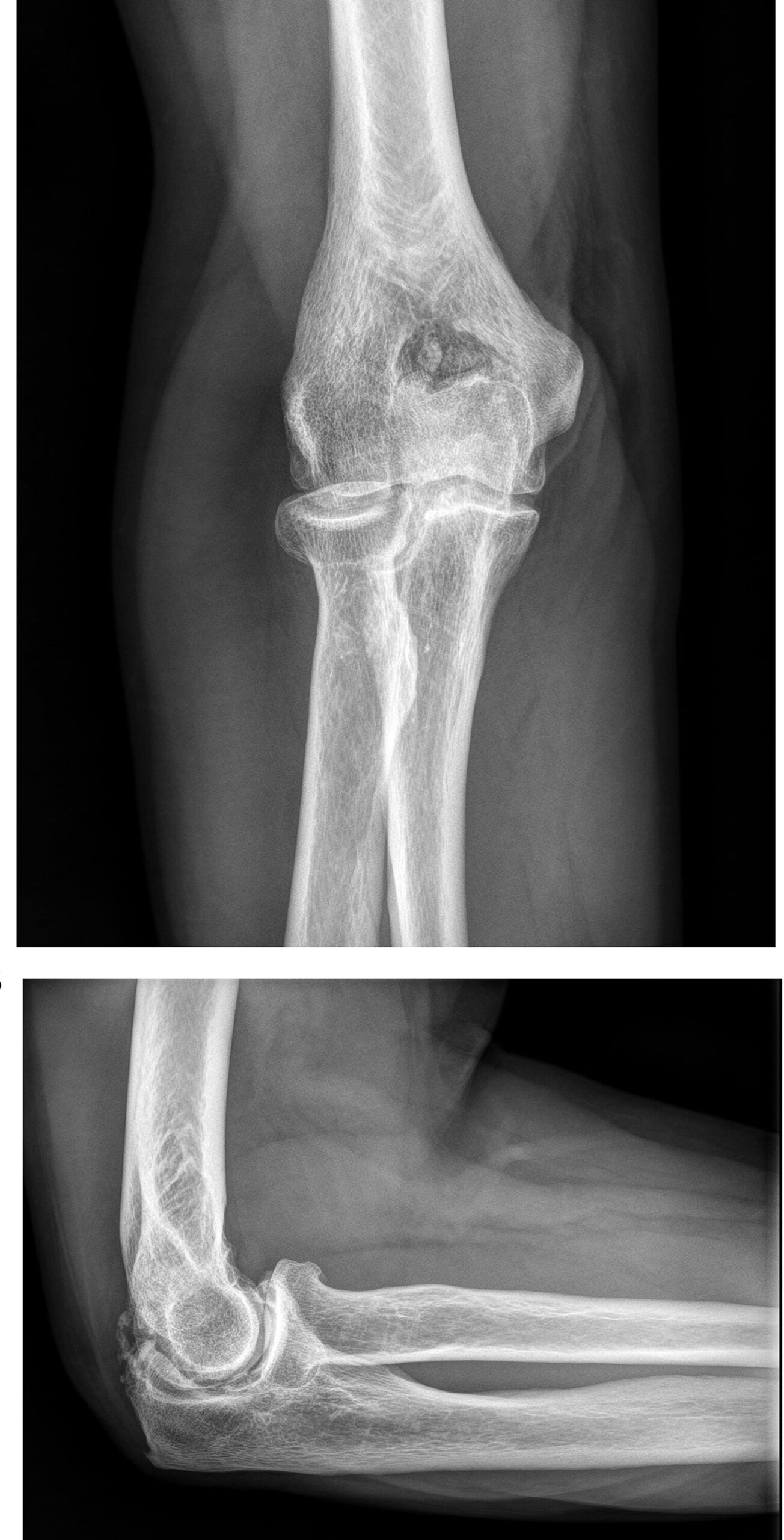
Right elbow extension is reduced (as pictured), with a hard end point and no ligamentous instability.
Flexion, supination and pronation are preserved and there is no bogginess or tenderness to palpation. There is no other joint involvement.
What is the most likely diagnosis?
Correct!
The findings are consistent with elbow arthritis, which is most commonly seen in the setting of rheumatoid arthritis, post-traumatic arthritis and primary osteoarthritis (OA).
Reduced range of motion with a hard end point suggests osteophytic impingement, compared with a soft end point, which is more likely to reflect soft tissue contracture or effusion
In this case the clinical picture is most consistent with primary OA, which affects around 2% of the population. It is most commonly seen in middle-aged to older men, affecting the dominant arm, typically in those with high-impact or repetitive-use occupations or sports, such as labourers, those engaged in throwing sports, or weightlifting.
Rheumatoid arthritis is an important differential diagnosis to consider, with elbow involvement reported in up to 61% of patients with elbow arthritis. Hallmark features include stiffness, synovitis with boggy tenderness and subchondral destruction, ultimately leading to medial and lateral collateral ligament deterioration and elbow instability.
Gout may be associated with olecranon bursitis, but typically this is seen with gout recurrences, and usually in the presence of other joint involvement (characteristically lower limb).
Septic arthritis is less likely given the patient is systemically well, the chronicity, and lack of local inflammatory features.
X-ray is a useful initial diagnostic examination, and may demonstrate peripheral osteophytes affecting the olecranon process, humeral fossae and coronoid, and loose bodies, resulting from wear of the articular surface. CT is the imaging of choice to see the actual bony loose bodies and to define the arthritis. MRI is not as useful as it does not define the bone changes as well as CT.
Typical non-surgical options for OA (simple analgesia, activity modification, corticosteroids) may be suitable for mild-moderate cases or in those with reasonably preserved function.
However, for patients with severe OA, with significantly impacted function or range of motion, surgery may be warranted. Options include arthroscopic elbow synovectomy and removal of the inevitable loose bodies or total elbow arthroplasty.
Younger patients are far more likely to benefit from an arthroscopic approach to clear the loose bodies but ultimately this will not reverse the early arthritic process.