
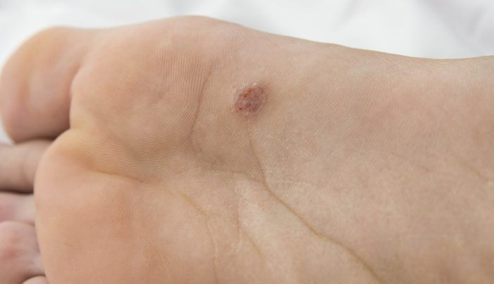
Hue is a 56-year-old executive of Chinese descent who presents with an enlarging lesion on his foot.
He first noticed a small, pink, raised, itchy nodule three months ago, and self-treated with home administered cryotherapy.
When the lesion kept enlarging, he tried some topical antibiotics left over from his child’s recent impetigo.
That was three weeks ago, and the lesion continues to enlarge and become more uncomfortable. Hue is otherwise well with no history of note.
Dermatoscopically it demonstrates asymmetrical variegated pigmentation from white to blue-grey. There is no lymphadenopathy.
What is the diagnosis?
Correct!
Acral lentiginous melanoma is the most common type of melanoma occurring in people with skin of colour, accounting for 40–60% of melanoma diagnoses in Asian and African-American ethnicities.
This was the suspected diagnosis in this case; however, after excisional biopsy confirmed malignant melanoma, subsequent wide surgical excision and immunohistochemical staining found this to be a primary nodular melanoma, with no locoregional spread.
Approximately 50% of melanomas arising at acral sites are acral lentiginous melanoma. Nodular melanoma accounts for 8-20% of acral lesions, and superficial spreading melanoma accounts for 35-40% of lesions.
In this case, the rapidity and ongoing growth of the lesion were clinical clues to the histopathology.
Rapid growth is a telltale feature of nodular melanoma, typically presenting as a lump that enlarges quickly over weeks to months.
Up to one-third of nodular melanomas are not pigmented. Other suggestive features include size >6mm, domed shape, single colour or variable pigmentation, ulceration, bleeding, itching or stinging.
Dermatoscopic clues include disorganised asymmetrical structure, atypical vascular patterns, blue-grey structures and multiple colours.
Differential diagnoses for amelanotic or hypomelanotic acral lesions include benign diseases such as warts, calluses or eccrine poroma (benign growths derived from cells of the terminal duct and connected to the epidermis.)
Pigmented actinic keratoses and seborrhoeic keratoses do not occur on acral surfaces.