
A 40-year-old woman of Iranian heritage presents to a dermatologist with a persistent, scaly, pruritic rash on her feet that she has had for many years. There is no rash elsewhere on her body.
She has previously been diagnosed with palmoplantar psoriasis and prescribed topical steroid creams, including betamethasone and oral prednisone. She also received ultraviolet light treatment. There was no improvement with any of these treatments.
She has no known allergies. Her cousin, sister and uncle have psoriasis. She lives at home with her husband and children and has a pet dog.
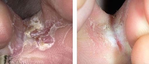
On examination, there is a well-demarcated erythematous rash on both, more marked on the left foot. This is associated with a yellowed, dysmorphic right toenail.
What is the most likely diagnosis?
Correct!
In this case, fungal microscopy and biopsy of the lesion confirmed Trichophyton species, consistent with a dermatophyte fungal infection. Tinea pedis is most commonly caused by T. rubrum, T. interdigitale and Epidermophyton floccosum dermatophyte fungi.
The condition typically presents as round, scaly, asymmetrical erosions with a well-defined edge. The rash is usually more inflamed on the edge compared to the centre. The dysmorphic, yellowed toenail is also suggestive of a fungal infection. This may be caused by a variety of factors, including humidity and occlusive footwear. Diagnosis is usually clinical but ideally confirmed with skin scrapings for microscopy and culture.
This patient had been incorrectly diagnosed with and treated for palmoplantar psoriasis for over two years. Palmoplantar psoriasis is characterised by scaly erythematous plaques that predominately affect the palms and soles. However, the prominent scaly edge and presence on the dorsal feet, seen in this case, is unusual for palmoplantar psoriasis. Additionally, the persistence and worsening of the rash, despite escalating treatment for psoriasis, further indicates this is not the correct diagnosis.
Discoid eczema is a common type of dermatitis that typically presents as round, erythematous, dry plaques that are mostly 1-3cm in diameter. The plaques are usually disseminated, typically including involvement of the arms and legs, and are extremely pruritic. Isolated involvement of the feet is not common. This type of dermatitis is associated with skin injury such as friction. Topical steroids and UV therapy are usually effective treatment options for this condition.
Granuloma annulare is characterised by smooth, round papules and plaques. It is ring shaped and usually localised to the hands and feet. Granuloma annulare is not scaly, and the centre of each ring is usually slightly depressed and clear in the middle.
This patient was treated with topical clotrimazole and oral itraconazole for three days, followed by topical terbinafine for three months. She was advised to keep her feet clean and dry. At review six weeks later, the rash had completely resolved.