
Ken, 19 years old, attends complaining of several weeks’ history of recurrent left facial swelling over the left angle of the mandible. The swelling lasts for about a week before resolving and is not painful. Ken denies any fever or systemic symptoms. He does note that his left third molar is erupting. His medical history is unremarkable, and he does not have any known allergies. He has never smoked and does not consume alcohol regularly.
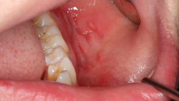
Extra-oral examination demonstrates a mild soft swelling over the left angle of the mandible. There is no erythema, and the area is not hot. Intra-oral examination demonstrates deep linear ulcers along the left buccal sulcus and gingival tissues of the left mandibular first and second molars. The left third molar is partially erupted, and the operculum does not appear to be inflamed. CT of the mandible does not demonstrate any pericoronal or periapical pathology associated with the third molar, but a diffuse non-specific soft tissue swelling is noted in the buccal sulcus and mucosa adjacent to the left mandibular molars.
What is the most likely diagnosis?
Correct!
Orofacial granulomatosis (OFG) is a rare chronic inflammatory and granulomatous condition. It may be idiopathic or present with other granulomatous diseases, including Crohn’s disease or sarcoidosis. OFG may occur at any age but is most common in young adults, without a sex predilection. Clinically, OFG presents as recurrent, usually painless soft orofacial swelling (most commonly the lip) which eventually becomes persistent and indurated, and may be aesthetically disfiguring. Other orofacial manifestations include an oral ulceration, gingival involvement, angular cheilitis and vertical fissures in established lesion.
Biopsy of an appropriate location and depth should demonstrate non-necrotising granulomatous inflammation, and the pathologist must exclude infectious causes (eg, mycobacterial or fungal infections) or foreign-body reactions. Investigation of systemic granulomatous diseases may include full blood count, haematinics, C-reactive protein, erythrocyte sedimentation rate, serum angiotensin converting enzyme level, ASCA and ANCA. If sarcoidosis is suspected, chest radiography may demonstrate bilateral hilar lymphadenopathy. As 10-40% of patients with OFG have associated Crohn’s disease, faecal calprotectin is useful, and referral to a gastroenterologist prudent even in the absence of gastrointestinal symptoms.
The management of OFG is often difficult, with variable and unpredictable clinical outcomes. Treatment may include dietary restrictions (eg, cinnamon- and benzoate-free diet), corticosteroid therapy (topical, intralesional and/or systemic) or other immunomodulatory drugs. There is insufficient evidence to support one therapeutic alternative over another.
A delay in diagnosis may result in firm, indurated and cosmetically undesirable orofacial swelling. However, when diagnosed and treated early, persistent orofacial swelling may be prevented. Follow-up is imperative, as recurrence is common.