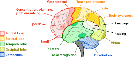
The neurological examination is a fundamental component of the overall physical assessment in healthcare, providing essential information about the nervous system’s functioning. It’s typically conducted when a patient presents with symptoms suggestive of a neurological disorder. The examination encompasses several steps, each designed to evaluate different aspects of nervous system function:
- Mental Status Examination: This part of the exam assesses cognitive functions including orientation (to time, place, and person), attention, memory, language abilities, and other higher cortical functions. It provides insight into the patient’s mood, thought content, and perception.
- Cranial Nerve Examination: The twelve cranial nerves are assessed to identify any abnormalities:
- evaluating the sense of smell (I),
- visual acuity and visual fields (II),
- eye movements (III, IV, VI),
- facial sensation (V),
- facial muscle movement (VII),
- hearing and balance (VIII),
- swallowing and palate elevation (IX, X),
- shoulder shrug (XI), and
- tongue movement (XII).
- Sensory Examination: This involves checking for the sensation of pain, temperature, vibration, and proprioception (sense of body position). Sensory testing can help localize lesions to specific neural pathways.
- Motor Examination: Involves assessing muscle tone, strength, and the presence of any involuntary movements. Observing the patient’s gait and posture can also provide valuable information about their motor system.
- Reflex Examination: Testing deep tendon reflexes (like the knee-jerk reflex) and superficial or cutaneous reflexes. Abnormal reflexes can indicate a problem in the reflex arc, including the sensory nerve, spinal cord, motor nerve, or muscle.
- Coordination and Gait Testing: This test assesses cerebellar function. It includes tests for coordination (such as the finger-to-nose test), rapid alternating movements, and gait assessment. Observing the patient’s walk can reveal problems with balance, coordination, or weakness.
- Special Tests: Depending on the patient’s symptoms and the findings of the above examinations, special tests may be performed
Special Tests
- Romberg Test:
- This test is used to assess proprioception and balance.
- The patient is asked to stand with feet together and arms at the side, first with eyes open and then closed.
- A positive Romberg test, in which the patient becomes significantly more unsteady with eyes closed, suggests a proprioceptive or vestibular deficit.
- Babinski Sign:
- To assess the integrity of the corticospinal tract.
- The examiner strokes the lateral aspect of the sole of the foot with a blunt object from heel to toe.
- An upward response of the big toe (dorsiflexion) with fanning of the other toes, which is known as a positive Babinski sign, is abnormal in adults and indicates central nervous system pathology, particularly in the corticospinal tract.
- Finger-Nose Test:
- This test is for cerebellar function.
- The patient is asked to touch their nose with their finger, then reach out to touch the examiner’s finger and go back and forth. Ataxia, characterized by a tremor during the movement, suggests cerebellar dysfunction.
- Heel-Shin Test:
- Another test for cerebellar function is where the patient is asked to slide their heel up and down the opposite shin.
- Inaccuracy in performing this task may indicate cerebellar ataxia.
- Kernig’s Sign:
- Used in the diagnosis of meningitis.
- While the patient is lying supine, the hip is flexed to a 90-degree angle, followed by an attempt to straighten the knee.
- Pain and resistance in the lower back and stiffness in the hamstrings represent a positive Kernig’s sign.
- Brudzinski’s Sign:
- Also used in meningitis diagnosis.
- The examiner flexes the patient’s neck; a positive sign is involuntary flexion of the hips and knees in response to this maneuver.
- Tandem Walking:
- Assesses gait and balance.
- The patient is asked to walk in a straight line by placing one foot directly in front of the other, heel to toe.
- Difficulty in performing this task can indicate problems with balance or cerebellar dysfunction.
- Graphesthesia:
- The ability to recognize writing on the skin purely by the sensation of touch.
- The inability to identify numbers or letters traced on the palm can indicate lesions in the sensory cortex.
- Stereognosis:
- This assesses the parietal lobe’s ability to integrate sensory information.
- The patient is asked to identify objects placed in their hand without visual input.
- The inability to recognize common objects by touch may suggest a parietal lobe lesion.
- Two-Point Discrimination:
- This test evaluates the patient’s ability to distinguish between one and two points when they are simultaneously applied to the skin.
- It is used to assess sensory nerve function and can indicate conditions affecting the dorsal columns of the spinal cord or the sensory cortex.
A thorough neurological examination helps localize the neurological lesion to a particular part of the nervous system, aiding in diagnosis and guiding further testing and management. As neurological conditions are quite common in the general population, it is crucial for healthcare professionals, particularly in general practice, to be proficient in conducting a neurological examination and interpreting its findings.
Cranial Nerves
The human body has twelve cranial nerves that are responsible for a variety of functions, including sensory and motor activities. Here is a list of the cranial nerves along with their primary functions:
- Olfactory Nerve (I):
- Function: Sense of smell.
- Type: Sensory.
- Optic Nerve (II):
- Function: Vision.
- Type: Sensory.
- Oculomotor Nerve (III):
- Function: Eye movement, opening of eyelid, pupil constriction, focusing.
- Type: Motor.
- Trochlear Nerve (IV):
- Function: Eye movement (specifically, the superior oblique muscle).
- Type: Motor.
- Trigeminal Nerve (V):
- Function: Sensation in the face and motor functions such as biting and chewing.
- Type: Both sensory and motor.
- Branches: Ophthalmic (V1), Maxillary (V2), Mandibular (V3).
- Abducens Nerve (VI):
- Function: Eye movement (specifically, the lateral rectus muscle).
- Type: Motor.
- Facial Nerve (VII):
- Function: Facial expressions, secretion of saliva and tears, taste (anterior two-thirds of the tongue).
- Type: Both sensory and motor.
- Vestibulocochlear Nerve (VIII):
- Function: Hearing and balance.
- Type: Sensory.
- Branches: Cochlear nerve (hearing), Vestibular nerve (balance).
- Glossopharyngeal Nerve (IX):
- Function: Taste (posterior one-third of the tongue), swallowing, secretion of saliva, monitoring blood pressure and oxygen levels in blood.
- Type: Both sensory and motor.
- Vagus Nerve (X):
- Function: Control of the heart, lungs, and digestive tract, sensation from the throat, and control of the muscles for speech.
- Type: Both sensory and motor.
- Accessory Nerve (XI):
- Function: Movement of the trapezius and sternocleidomastoid muscles (shoulder shrugging and head turning).
- Type: Motor.
- Hypoglossal Nerve (XII):
- Function: Tongue movements.
- Type: Motor.
These cranial nerves are integral to a wide range of functions, from the sensory perception of the external environment to motor control of muscles and glands.