
Ali, a 19-year-old university student, presents for review of progressive thinning of his eyebrows.
There is associated facial erythema and ‘bumpy spots’ on the forehead and upper arms, present since adolescence.
He has previously been diagnosed with rosacea and acne, and has trialled topical and oral antibiotics, with no benefit. He currently uses a simple fragrance-free cleanser only.
Ali is otherwise well, with no regular medications and no systemic features of note.
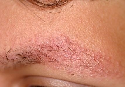
On examination, the eyebrows show areas of alopecia, with well-defined confluent follicular erythematous papules.
There are multiple, well-defined red to brown follicular papules affecting the forehead and upper arms bilaterally.
He has central facial erythema with nasolabial sparing. The scalp and beard are not affected.
Blood tests are normal, including inflammatory markers, vitamin B12, iron and thyroid function.
What is the most likely diagnosis?
Correct!
Keratosis pilaris rubra atrophicans faciei is a rare form of keratosis pilaris, characterised by perifollicular erythema and keratosis papules involving the malar region, forehead, temples and eyebrows.
Scar-like follicular depressions result, leading to atrophy and loss of hair in the affected area.
The periauricular regions of the face and scalp may also be affected, and there is often associated keratosis pilaris of the upper arms.
Cases are typically sporadic. Rarely, they are attributable to autosomal recessive mutations in the desmoglein 4 gene, or associated with congenital syndromes such as Noonan syndrome.
The pathogenesis is of abnormal follicular keratinisation, causing scale to fill the follicle and obstruct the hair shaft, resulting in inflammation. Over time, this leads to atrophy and alopecia.
The diagnosis is clinical. Biopsy, if performed, reveals nonspecific histopathologic features including a widened infundibulum, plugged hair follicle, loss of hair follicle, superficial perivascular lymphocytic infiltrate, and dermal fibrosis.
Treatment aims to minimise follicular scale and erythema. Scale may be managed with soap-free cleansers, exfoliation with a pumice or sponge, keratolytic moisturisers and topical retinoids or calcineurin inhibitors.
Sun protection is recommended to reduce erythema, and pulse dye laser or intense pulsed light may also be of benefit. The features of this condition tend to regress naturally after puberty; however, any associated atrophy and hair loss is irreversible.
Phrynoderma is a form of minimally symptomatic follicular hyperkeratosis, in the setting of nutritional deficiency. The back of the elbows and front of the knees are first affected, with subsequent spread to the extremities, upper forearms and thighs. The face is rarely involved.
Frontal fibrosing alopecia is a form of follicular lichen planus that results in loss of lateral eyebrows and frontal hairline due to scarring alopecia, as a result of facial follicular papules. This is mainly seen in postmenopausal women.
Darier disease causes scaly papules on the hands and seborrhoeic areas, including the nasolabial folds, forehead, scalp and ears.