X-ray rules refer to the guidelines and principles used by healthcare professionals to determine when to order an X-ray and how to interpret the results. These rules help in making effective and efficient use of radiographic imaging in various clinical scenarios. Let’s discuss some key aspects:
Indications for Ordering an X-ray
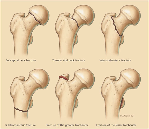
- Trauma: To evaluate bone fractures, dislocations, or joint injuries.
- Infection: Suspected osteomyelitis or septic arthritis.
- Chronic Pain: To investigate potential causes like arthritis or bone deformities.
- Respiratory Symptoms: For suspected pneumonia, lung tumors, or other chest pathologies.
- Abdominal Pain: To detect obstructions, perforations, or certain abdominal pathologies.
- Foreign Body Detection: In cases of suspected ingestion or insertion of foreign objects.
- Postoperative Evaluation: To assess the placement of surgical hardware or the status of a healing bone.
Basic Interpretation Principles
- Patient Identity
- Adequacy and Quality of X-ray: Ensure the X-ray is of good quality and shows the area of interest adequately.
- Systematic Approach: Always follow a systematic approach to avoid missing key findings ie ABCDEFGHI approach to CXR
- Comparison with Previous X-rays: Helps in identifying new changes or the progression of a condition.
- Look for the Obvious and Then the Subtle: Identify obvious abnormalities first, then focus on more subtle details.
Specific Rules and Guidelines
- Ottawa Ankle and Foot Rules: Used to decide if an X-ray is needed for ankle and foot injuries.
- Ottawa Knee Rules: Guidelines for when to X-ray a knee after an injury.
- C-spine Rules: To determine the need for cervical spine X-ray in trauma cases.
- ABCDs of Bone X-ray Evaluation: Assess Alignment, Bone density, Cartilage spaces, and Soft tissues.
- FABERE/Patrick’s Test for Hip Pathology: A clinical test that, if positive, may warrant an X-ray. Flex/Abduct/Ext Rotate (like yoga lotus position)
- ABCDEFGHI CXR Approach: Assess the airway, bones and soft tissues, cardiac, diaphragms, equal volume, fine detail, gas under diaphragm, hilum, ID and impression
Safety and Regulatory Considerations
- Minimizing Radiation Exposure: Especially important in children and pregnant women.
- Proper Shielding: Using lead aprons or other protective measures when necessary.
- Legal and Ethical Guidelines: Following appropriate guidelines for the use and interpretation of X-rays.
Conclusion
The decision to order an X-ray and the interpretation of its findings should be guided by clinical judgment, patient history, physical examination, and specific clinical rules. These rules help in making judicious use of radiographic imaging, reducing unnecessary exposure to radiation, and ensuring accurate diagnosis and management.
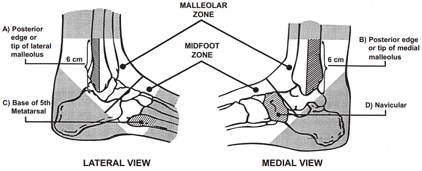
Ottawa Ankle and Foot Rules
These rules are designed to minimize unnecessary ankle and foot X-rays. They suggest an X-ray is only needed if there is pain in the malleolar or midfoot zone and any one of the following:
- Ankle Rules:
- Bone tenderness at the posterior edge or tip of the lateral or medial malleolus.
- Inability to bear weight both immediately and in the emergency department (four steps).
- Foot Rules:
- Bone tenderness at the base of the fifth metatarsal (for midfoot injuries).
- Bone tenderness at the navicular bone.
- Inability to bear weight both immediately and in the emergency department.
Ottawa Knee Rules
These rules are used to determine when an X-ray is necessary for knee injury patients. An X-ray is indicated if the patient has:
- Age 55 or older.
- Isolated tenderness of the patella (with no other bone tenderness in the knee).
- Tenderness at the head of the fibula.
- Inability to flex the knee to 90 degrees.
- Inability to bear weight both immediately and in the emergency department (four steps).
Canadian C-spine Rules
These rules help decide whether cervical spine X-rays are necessary for trauma patients. X-rays are required if any of the following criteria are met:
- Age 65 or older.
- A dangerous mechanism of injury (e.g., fall from a height, motor vehicle accident).
- Paresthesias in the extremities.
- Inability to actively rotate the neck 45 degrees to the left and right.
ABCDs of Bone X-ray Evaluation
This systematic approach is used for evaluating bone X-rays:
- A for Alignment: Look at the general architecture of the bones.
- B for Bone density: Examine the bone density for signs of osteopenia or focal lesions.
- C for Cartilage spaces: Check the joint spaces to see if they are narrowed, which might suggest arthritis.
- D for Soft tissues: Look for any soft tissue abnormalities or swelling.
FABERE/Patrick’s Test for Hip Pathology
While not a rule for X-ray per se, the FABERE (Flexion, ABduction, External Rotation, and Extension) test can indicate if a hip X-ray is necessary:
The patient lies on their back, and the clinician flexes, abducts, and externally rotates the patient’s hip. Pain or discomfort may suggest the need for further investigation, possibly including an X-ray, especially if clinical suspicion for a hip pathology is high.
Applying These Rules
These rules are guides and not absolutes. Clinical judgment is crucial, and the rules should be applied in the context of the patient’s overall clinical picture. If there’s uncertainty, or if symptoms persist despite negative initial X-rays, further evaluation may be warranted.
ABCDEFGHI CXR
The ABCDEFGHI approach is a systematic method for evaluating a chest X-ray (CXR) to ensure a thorough examination and to minimize the risk of missing important findings. Each letter corresponds to a specific aspect of the CXR to check:
- A – Airway
- Trachea: Check the position of the trachea; it should be central or slightly deviated to the right.
- Carina: Look at the carina and the angle it makes.
- Main Bronchi: Observe the main bronchi for any abnormalities or deviations.
- B – Bone and Soft Tissues
- Bones: Inspect the bones (ribs, clavicles, spine) for fractures or lesions.
- Soft Tissues: Look at the soft tissues around the thorax for masses or subcutaneous emphysema.
- C – Cardiac Silhouette
- Size and Shape: Assess the size and shape of the heart. The heart should occupy less than half the width of the thorax.
- Borders: Identify the borders of the heart and note any irregularities or enlargements.
- D – Diaphragm
- Level: Check the diaphragm’s level (right usually higher than left)
- Contour: Flattened diaphraghms are seen in the hyperinflation of COPD.
- Costophrenic Angles: Look for blunting which could indicate pleural effusion.
- E – Equal Volume (Look for pneumothorax)
- Pneumothrax: This can be very obvious or quite subtle and only seen at the lung apex
- F – Fields
- Lung Fields: Examine for any abnormalities like consolidation, atelectasis, pneumothorax, or masses.
- Texture and Markings: Look for changes in lung markings or texture, indicating conditions like fibrosis, edema, or chronic obstructive pulmonary disease (COPD).
- Pleural Fluid: Look for fluid in the pleural spaces, which may appear as dense shadowing at the lung bases.
- G – Gas Under The Diaphragm
- Gastric Bubble: Ensure the gastric bubble is present and located under the left hemidiaphragm.
- Free Gas: Can be any size and position under the diaphragm but has a thin line of diaphragm above.
- H – Hilum and Mediastinum
- Hilar Structures: Evaluate the size and position of the hilar shadows.
- Mediastinum: Examine the width and contents of the mediastinum for masses, lymphadenopathy, or widening that could suggest aortic aneurysm.
- I – Identity and Impression
- Indentity: Make sure you are looking at the correct patient’s Xray!
- Clinical Impression: Correlate the radiographic findings with the patient’s clinical presentation and history.
This ABCDEFGHI approach ensures a comprehensive evaluation of a chest X-ray, covering all major anatomical and pathological areas of interest. It’s important for clinicians to be methodical in their analysis to avoid missing subtle yet clinically significant findings.